what is a 3t mri used for
This is why you get the same resulting output image as on a real scanner given the same inputs being used including anatomy resolution scan time SNR contrast and artifacts. MRI does not involve X-rays or the use of ionizing radiation which distinguishes it from.
Mri Machines Understanding The Differences
With that said a 3T machine isnt always the best for every kind of imaging.

. ImageReady MR Conditional Pacing System as the system is designated as MR Conditional in accordance with specific conditions. MRI scanners use strong magnetic fields magnetic field gradients and radio waves to generate images of the organs in the body. Spinal Fixation Hardware The main issue with imaging spinal fixation hardware including Harrington rods is artifact.
Magnetic resonance imaging is one of the most informative diagnostic techniques. 1-4 Historically less than 1 would have received a scan. The FDA website has more information.
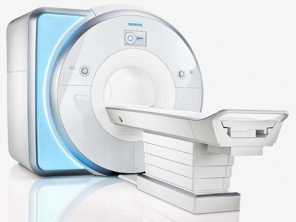
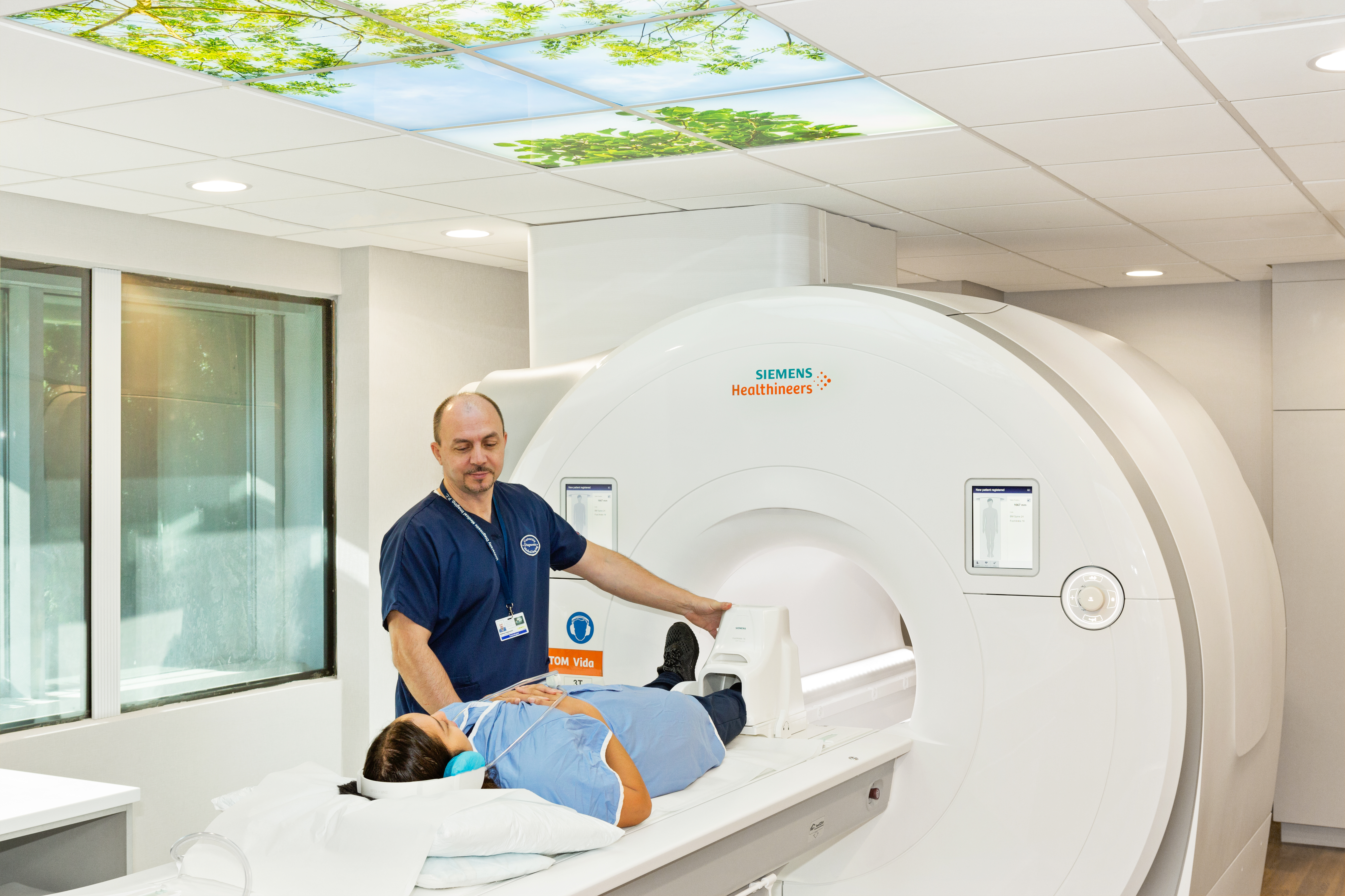
Chronotropic competence is defined by the Model of the Cardiac Chronotropic Response to Exercise. MAGNETOM Lumina is the new 3T Open Bore system that gives you full confidence to deliver the productivity reproducibility and patient satisfaction that you demand in MRI. PC-MRI may be considered a method of magnetic resonance velocimetry.
MRI is an abbreviation for magnetic resonance imaging. The majority of MRI systems in clinical use are between 15T and 3T. 1-4 Today with our exclusive MRI portfolio you can ensure safe MRI access for most patients when MR conditions are met.
Magnetic resonance imaging MRI is a medical imaging technique used in radiology to form pictures of the anatomy and the physiological processes of the body. Magnetic resonance imaging MRI has become the premier orthopedic diagnostic tool used in detecting meniscal and anterior cruciate ligament ACL tears and has virtually replaced both arthrography and arthroscopy as the diagnostic test of choice. An MRI scan provides higher resolution images and better soft tissue contrast.
We used the commonly used weightings and RFP-FatSat T1 and impacts axial coronal and sagittal in medical imaging studies of the hip. The 3T WBMR images were acquired in a head-to-toe scan in 20 patients with RA and at least 1. Annually 1216 of device patients are likely to have an MRI ordered.
Both types currently pose hazards to patients referred to MRI procedures. To obtain magnetic resonance imaging scan an MRI scanner is used. There are two basic types of insulin pumps one is used as an external device and the other is implanted.
Our technology is built on more than 8 years of research on MRI simulations. Medtronic has long been committed to providing cardiac device patients with access to the. The strength of electromagnets used to pick up cars in junk yards is about the field strength of MRI machines 15 to 20T.
Phase contrast MRI PC-MRI is used to measure flow velocities in the body. Powered by our premium MR technology MAGNETOM Lumina combines our unique BioMatrix technology the new syngo MR XA software platform and our exclusive Turbo Suite to. The MRI images of the elbow derived from a healthy volunteer.
Roger Woods Center Director x44057 5. This type of scan uses magnetic fields and radio waves in a non-invasive way to diagnose problems inside the body. It is used mainly to measure blood flow in the heart and throughout the body.
Image acquisition was performed on a 3 Tesla MRI with a thickness of 4 mm in contiguous sections. The StandUp MRI Offers Extraordinary Patient Comfort and Diagnostic AdvantagesThe unique physical configuration of the StandUp MRI makes it the worlds most patient-friendly MRI and also the only MRI that can scan patients in any position including weight-bearing positions allowing it to detect problems that would be underestimated or completely missed by any other. They have a wide range of applications.
Since modern PC-MRI typically is time-resolved it also may be referred to as 4-D imaging three spatial dimensions plus time. The B o in MRI refers to the main magnetic field and is measured in Tesla T. Trent Thixton Lead MRI Tech x59217 4.
BMC Techs BMCTechsmednetuclaedu 424 652-6290 6. It serves as the definitive resource for radiologists and other physicians MRI technologists physicists. 3T has a higher likely hood of artifacts.
The commonly used Corflo feeding tube the yellow tube including those with tungsten-weighted tips is MRI safe at 15T and 3T with the stylet removed. MRI BIOEFFECTS SAFETY AND PATIENT MANAGEMENT is a comprehensive authoritative textbook on the health and safety concerns of MRI technology that contains contributions from more than forty internationally respected experts in the field. Traditional MRI machines operate at a strength of 15T or Teslas.
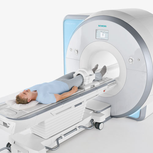
A wide bore MRI features all of the benefits of a closed bore MRI with more space inside the bore area of the machine. 3T is ideal for imaging small bones breast MRI musculoskeletal MRI neurological MRI and vascular MRI where the minute details are especially crucial to diagnosis. Inside each MRI scanner is a very large magnet of varying strength.
The earths magnetic field is about 05 gauss. This non-invasive procedure is absolutely painless and provides maximum useful information. BMC Emergency ONLY Line.
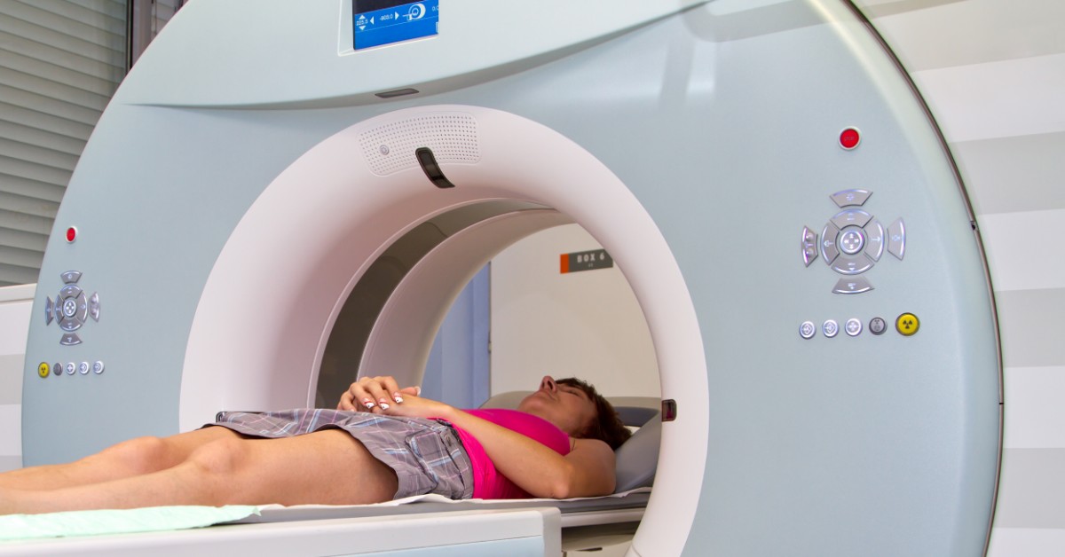
A tesla is the unit of measurement quantifying the strength of a magnetic field. It produces detailed images of organs bones and soft tissue. Its popularity is explained by the highest accuracy comfort and simplicity.
We offer several different types of short bore MRI machines at our various locations across New Jersey. A short bore MRI enables more of your body to remain outside of the MRI machine specifically your head. These different strength magnets are measured in Teslas or T.
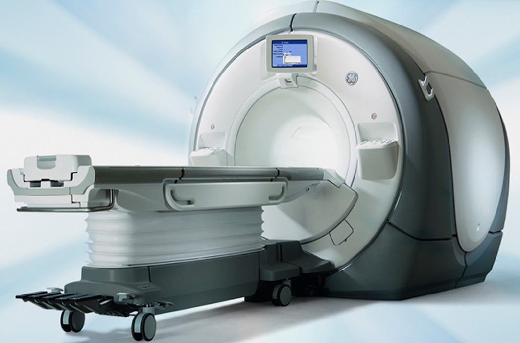
A 3T MRI generates a magnetic field that is twice as strong as a normal MRI and 10 to 15 times as strong as open MRI scanners. Please refer to the MRI Technical Guide. An MRI scanner is a very large piece of diagnostic imaging equipment that is used in radiology and medical imaging to investigate the anatomy and physiology of the body.
This implant is totally contraindicated for MRI in both the 15T and 3T. Mary Susselman PETMRI Tech x64291 3. For an external insulin pump in general.

3 T 70 Cm Philips Ingenia Elition X Advanced Magnetic Resonance Imaging And Spectroscopy Amris Facility Mcknight Brain Institute University Of Florida
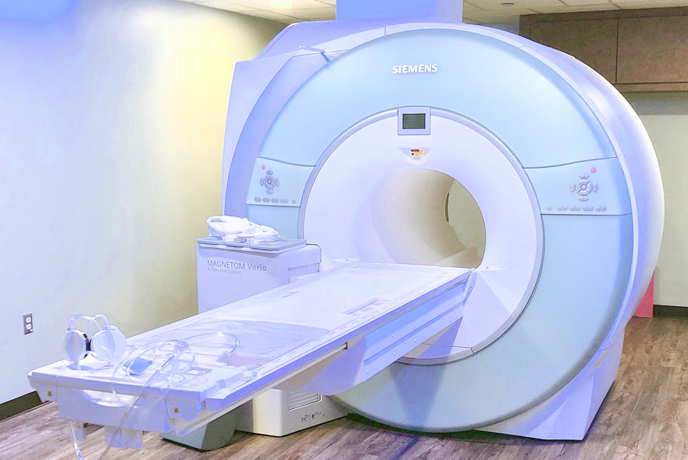
Mri Scan Siemens 3t Skyra Scanner Advanced Imaging Of South Bay Torrance San Pedro Califonria
Optimal Imaging Jacksonville Mri

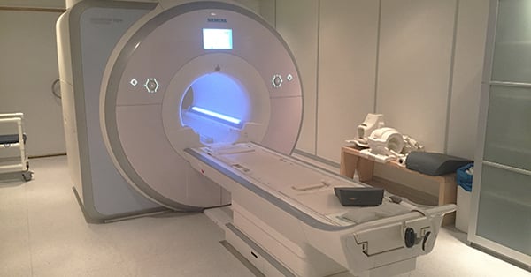
Siemens Magnetom Skyra 3t Mri Machine References Radiology Kovai Download Scientific Diagram

Do You Need To Prepare For An Mri Scan Medical Associates Of Northwest Arkansas

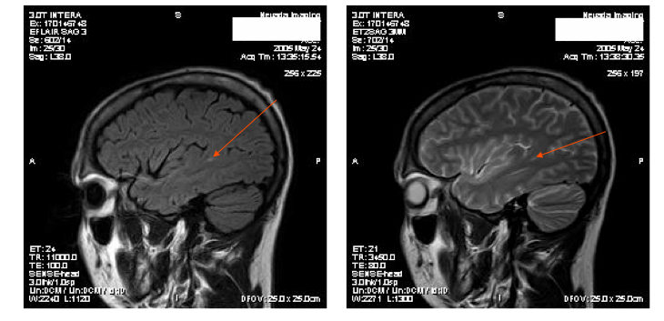
1 5t Versus 3t Mri

Difference Between A 1 5t And A 3t Mri Scanner Omega Pds
3t High Resolution Mri Vs Normal Mri Affinity Radiology
Differences Between 3t And 1 5t Mri Machines
3t Magnetic Resonance Imaging Mri Santa Fe Imaging
3t Mri Vs 1 5t Mri

3t Mri Scanning Heath Lodge Clinic Cmc Imaging

Why The 3 Tesla Mri Is The Best Scanner For Diagnostic Imaging Rai Health Awareness Blog
3t Mri The Latest Advancement In Diagnostic Imaging University Diagnostic Medical Imaging

3 Tesla 3t Mri At Radiology Associates Of Ridgewood Radiology Associates Of Ridgewoodradiology Associates Of Ridgewood

Mri Ny Imaging Specialists
3t Mri Increased Field Strength To Image Mtbi
Mri Monash Biomedical Imaging

About Our 3t Mri Machine Omega Pds